THE HONG KONG MINIMALLY INVASIVE BRAIN & SPINE NEUROSURGERY CENTRE

 
NEUROSURGERY is a highly specialized surgical subspecialty that involves i) medical treatments ii) physical treatments iii) interventional or endovascular procedures and iv) minimally invasive microsurgeries for treating a very wide spectrum of clinical conditions and diseases that happen at our BRAIN and SPINE. Among all, treating STROKE and PAIN are the basics of Neurosurgery daily practices.

In the other words, NEUROSURGEONs, with their knowledges and trainings, together with their sophisticated micro-instruments, utilize their operative skills under the microscope, they treat wide spectrum of clinical conditions and diseases that happen along human nervous systems and its surrounding tissues; skull, spinal column, vessels and related tissues.

The followings are the summary of the common routines in Neurosurgical Practices:

  1. Head trauma and brain injury

  2. Brain tumors and Pituitary tumors:

  3. Stroke: both Ischemic and Hemorrhagic strokes

    1. Stroke Risk Assessment and Stroke Screening

    2. Stroke Prevention:

    3. Stroke Acute Treatment within Golden-Hours;

  4. Neuropathic Pain Treatments and Functional Neurosurgery:

    1. Trigeminal neuralgia
    2. Glossopharyngeal neuralgia
    3. Hemifacial spasm

    Microvascular Decompression (MVD)

  5. Spine:

    1. Spinal Tumor

    2. Spinal Trauma:
       with or without spinal fracture, spinal cord or nerves injury.

      1. Minimally invasive decompression +/- fixation spinal surgery for spine stabilization.

    3. Spinal Herniated Disc:

      with numbness, pain, weakness at arms or legs, sciatica pain at legs.

      1. Spinal injection and radiofrequency treatments for discogenic back pain or sciatica pain at legs.
      2. Microdiscectomy for freeing the impinged nerves
      3. Mini-decompressive Laminectomy for freeing the impinged spinal cord or nerves.

    4. Spinal Stenosis at neck or lower back:

      with spinal cord or cauda equina nerves compressions, causing pain, numbness, weakness at arms or legs, disturbed gait, disturbed urinary habit and bowel habits, disturbed sexual function.

    5. Degenerative Spine Disease:

      with back pain, nerves compressions, spinal instability or scoliosis: 

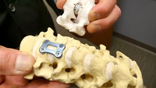
      Minimally Invasive Spine Fusion Surgery under Microscope:
      1. Transforaminal Lumbar Interbody Fusion (TLIF)
      2. ObliquLumbar Interbody Fusione (OLIF)

Among all Neurosurgeries, minimally invasive spine surgery (MIS Spine Surgery) are the microsurgeries that Neurosurgeons most commonly perform among their routines. 

For all spine surgeries, Neurosurgeons apply their knowledges of nervous system and spinal column, and also their microsurgical skills and techniques, together with their micro-instruments of brain surgeries. Sometimes, Neurosurgeons may also add the technique of Continuous Intra-Operative Neural Monitoring (IOM) to monitor the nerve signal transmission function along brain, spinal cord and nerves during surgery, so as to assure patients’ good neurological outcome in spine surgeries. 

Seeking Second Medical Opinion

With the ever-advancing Medical Technology, seeking a second medical opinion is now the standard practice for all kinds of medical diagnoses. Getting a second medical opinion from another Medical Expert is a decision-support tool for ratification or modification of the medical suggestions from other doctors.

Second medical opinion may have critical impacts on the disease outcome by influencing the followings, namely 5Cs:

Head injury

Brain tumors can be subdivided into non-cancerous benign tumor and malignant cancerous tumor. If a tumor is originated within brain compartment ......

Spine Surgery
Spine Surgery

Tumors can develop in the vertebrae, nerves, and other tissue throughout your spine. Some spine tumors, such as astrocytomas, occur more commonly in children and adolescents......

Stroke Prevention

Given the disease burden of strokes, prevention is an important public health concern. As stroke neurosurgeons, we do not want to treat stroke unless we are forced to do so for acute stroke ......

brain tumor treatment Hong Kong

Brain tumors grow and compress normal brain tissue. Both benign and malignant tumors can cause swelling of the brain and raised intracranial pressure. Headache, dizziness....

 
Interventional Pain Management


Pain is a multidimensional experience. Chronic pain differs from acute pain in that it lasts for more than 3-6 months, and there may not be obvious tissue injury leading to the pain. The pathway leading from stimulus to perception may be sensitised [1]. There is often associated depression. Management of chronic pain therefore requires a holistic multi-disciplinary approach [2]. In addition to pharmacological treatment, psychosocial support, physiotherapy and operative treatment, interventional techniques may benefit some patients by defining the pain generator and offers prolonged relief.

Interventional Pain Procedures
Commonly performed interventional procedures for pain of spinal origin include trigger point injections, facet blocks, sacroiliac joint blocks, epidural steroids and epidural lysis. Blocks with local anaesthetics identify the source of pain when pain level is reduced significantly after the block. Local administration of steroids decreases inflammation. Denervation by a radiofrequency current can bring prolonged relief by interrupting the sensory pathway. A very small group of selected patients benefits from spinal cord stimulation or insertion of an intra-thecal drug delivery system. Diagnostic blocks with a local anaesthetic help to ascertain the source of pain.

Back & Neck Pain
Facet joints are small synovial joint connecting the posterior aspects of the vertebrae. Inflammation of these joints or abnormal stress on them can lead to facet joint pain. There are back or neck pain with radiation, but no radicular symptoms. Pain typically worsens on extension. Facet pain syndrome often co-exists with disc pathologies [3]. These joints are supplied by medial branches of adjacent spinal segments. Diagnostic blocks are performed by injection of the joints or the medial branches under fluoroscopy. Joint injections can be supplemented by local steroids. Longer term effect is achieved by radiofrequency lesioning of the medial branches [4,5] (Fig.1).
 
    
Fig. 1a. Radiofrequency denervation of C2/C3 facet joint   Fig. 1b. Radiofrequency ablation of lower cervical medical branches
 
Sacro-Iliac Joint Pain
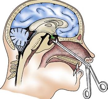
Sacroiliac joint pain presents with back pain, usually to the side of lesion, with radiation to the groin or the knee. Bilateral joint involvement may present as central back pain. Interventional treatment approach is similar to facet joint pain [6,7]. More recently developed Cooled Radiofrequency technique produces more consistent lesions for the multiple sacral nerves [8].

Sciatica
Radiculopathy presents with back or neck pain with radiation. Lesions in the lumbar levels produce the familiar ‘sciatica’ symptoms, whereas cervical lesions cause pain radiating to the shoulder or upper limb. Radicular pain may be caused by pressure or irritation of the nerve roots by degenerated intervertebral discs. Nerve impingement may also be caused by spinal stenosis, spondylolisthesis or failed back surgery syndrome. Injection of steroids into the epidural space may help these patients. The approaches may be translaminar, transforaminal or caudal [9,10] (Fig. 3). If multiple levels involvement is suspected or for resistant cases, a specially designed Racz catheter can be introduced from the sacral hiatus to define the levels of nerve impingement on epidurogram [11]. Hydrodissection is performed and steroid is deposited [12] (Fig. 4).
 
          
Fig. 3. Epidural lysis of adhesions   Fig. 4. Cervical transforaminal epidural steroid injection
 
Spinal Cord Stimulation
Melzack and Wall proposed the gate-control theory in 1965 [13]. The concept of stimulation of large afferent fibres to close the ‘gate’ for pain perception lead to the development of spinal cord stimulation. Shealy published the application of ‘dorsal column stimulation’ in 1967 [14]. Over the years spinal cord stimulation has developed into a percutaneous technique where a lead with multiple contact points is inserted into the epidural space and is connected to an implanted pulse generator. Programmable electric current stimulates the spinal cord, leading to pain relief. This is particularly useful for very carefully selected patients who suffer from failed back surgery syndrome [15] or arachnoiditi. (Fig.4)

Intrathecal Drug Delivery Systems
Implantable intrathecal drug delivery systems are available for delivery of medications such as opioids or baclofen into the subarachnoid space directly. This may be of use for some very selected patients [16].
 
 
References
  1. Latremoliere A, Woolf CJ. Central sensitization: a generator of pain hypersensitivity by central neural plasticity. J Pain 2009;10:895-926.
  2. Siddal PJ, Cousins MJ. Persistent pain as a disease entity: implications for clinical management. Anesth Analg 2004;99:510-20.
  3. Schwarzer AC et al. The relative contributions of the disc and zygapophyseal joint in chronic low back pain. Spine 1994;19:801–6.
  4. Boswell MV et al. A systematic review of therapeutic facet joint interventions in chronic spinal pain. Pain Physician 2007;10:229-53.
  5. Falco FJE et al. Systematic review of diagnostic utility and therapeutic effectiveness of cervical facet joint interventions. Pain Physician 2009;12:323-44.
  6. Cohen SP. Sacroiliac joint pain: a comprehensive review of anatomy, diagnosis, and treatment. Anesth Analg. 2005 Nov;101(5):1440-53.
  7. Rupert MP et al. Evaluation of sacroiliac joint interventions: a systematic appraisal of the literature. Pain Physician. 2009 Mar-Apr;12(2):399-418.
  8. Patel N et al. A randomized, placebo-controlled study to assess the efficacy of lateral branch neurotomy for chronic sacroiliac joint pain. Pain Medicine 2012; 13: 383–398.
  9. Parr AT et al. Lumbar interlaminar epidural injections in managing chronic low back and lower extremity pain: a systematic review. Pain Physician 2009; 12:163-188
  10. Gharibo CG et al. Interlaminar versus transforaminal epidural steroids for the treatment of subacute lumbar radicular pain: a randomized, blinded, prospective outcome sstudy. Pain Physician 2011; 14:499-511.
  11. Racz GB et al. Percutaneous lysis of epidural adhesions-evidence for safety and efficacy. Pain Pract. 2008 May 23.
  12. Epter R et al. Systematic review of percutaneous adhesiolysis and management of chronic low back pain in post lumbar surgery syndrome. Pain Physician. 2009; 12:361-378.
  13. Melzack R, Wall PD. Pain mechanisms: a new theory. Science 1965;150:971-9.
  14. Shealy CN, Mortimer JT. Electrical inhibition of pain by stimulation of dorsal columns: Preliminary clinical report. Anesth Analg 1967; 46: 489-91.
  15. North RB et al. Spinal cord stimulation vs reoperation for failed back surgery syndrome: A cost effectiveness and cost utility analysis based on a randomized controlled trial. Neurosurgery 2007; 61(2):361-8.
  16. Patel VB et al. Systematic review of intrathecal infusion systems for long-term management of chronic non-cancer pain. Pain Physician 2009; 12:345-360
The New Concept and the Latest Advances in Stroke Management
 
Summary
The old concept of 'Cerebrovascular Accident" is recently replaced by the new concept of "Brain Attack". Just like heart attack, brain attack is also an acute cardiovascular disease. Both heart attack and brain attack are of similar etiologies. The concept of treatment for heart attack also applies to brain attack. For heart attack, cardiologist prefers Percutaneous Coronary Intervention, the cardiac-PCI rather than intravenous recombinant tissue plasminogen activator (iv rtPA or 'Activase'). Cardiac-PCI has to be performed within 12 hours after symptom onset. IV rtPA is thus the 2nd line treatment for heart attack, offered only if PCI is not available.

Similar concept also applies to brain attack. Vascular Neurosurgeon also prefers Percutaneous Cerebral Intervention (Neuro-PCI), rather than iv rtPA. The Neuro-PCI has to be performed within 8 hours after symptom onset. This is thus the new Golden-8-hours Concept for brain attack. Nowadays Neuro-PCI is the first line standard treatment offered by most Comprehensive Stroke Centres in US. Iv rtPA within Golden-3-hours is considered to be the 2nd line treatment, offered only when Neuro-PCI is not available.


The procedures of mechanical thrombectomy


                        Fig 1 a - g. The procedures of mechanical thrombectomy

 
  a. Pretreatment angiography shows total occlusion of the M1 segment of the left middle cerebral artery (MCA) (arrow).
b  and c. Angiogram and illustration of the procedure show the occlusion site and surrounding angioarchitecture.
d. Immediate post-procedural angiography shows complete revascularisation (arrow).
e. Large syringe is connected to the reperfusion catheter with forceful suction.
f.  Disrupted clot removed out from body.
g. Retrieved whole clot


The New Concept of Stroke
The old concept of cerebrovascular accident and the acceptance of the fate of gradual neurological deterioration and the final state of disability, or even death, should have a revolutionary change. The new term "Brain Attack" implies that stroke is not an accident. Just like a heart attack, a stroke is something preventable, treatable, or even curable, without leaving patients with any neurological deficits. Strokes can be tackled in a similar way to heart attacks. In future, patients suffering from acute ischemic strokes will be less miserable. [1,2]
 
What is a stroke?
The term "stroke" refers to the sudden onset of neurological impairment due to vascular causes. There are two types of stroke: Ischaemic stroke accounts for ~87% of all strokes in the western world (~70% in Hong Kong). Haemorrhagic stroke explains the remaining 13% (~30% in Hong Kong). Yet haemorrhagic stroke has a higher mortality than the ischaemic entity; it is responsible for more than 30% of all stroke deaths.
 
Pathophysiology
Strokes can strike individuals regardless age, race or gender. During ischaemic stroke, a blood vessel in the brain becomes blocked. The blockage disrupts the blood flow, preventing oxygen from being delivered to the area of the brain affected. The section of brain tissue deprived of oxygen becomes "stunned" and no longer functions properly, resulting in the stroke symptoms.
 
Time is Brain
Acute ischaemic stroke (AIS) is a serious medical emergency. Rapid intervention is crucial. For every minute a large vessel stroke goes untreated, as many as 1.9 million neurons are lost (32,000 neurons are lost per second, 120 million neurons are lost per hour), 14 billion synapses are lost , 7.5 miles of myelinated fibers are lost. Permanent neurologic damage is more likely to occur the longer a stroke goes untreated. If treatment is delayed, additional death of critical brain cells occurs "infarction" reducing the potential benefit of the treatment. A short time to treatment is absolutely critical to increase the probability of a good recovery. After approximately three hours, brain cells will suffer irreversible injury usually leading to their death. This is the reason why thrombolytic therapy of rtPA are given only until three hours since the onset of the stroke. Therefore time really is brain.[3]
 
Epidemiology and Socio-Economic Impact
  1. Each year roughly there are 18,000 stroke cases in Hong Kong. Each year roughly 795,000 Americans experience a new (610,000) or recurrent (185,000) stroke.
  2. Stroke is the third leading cause of death in the United States, behind heart disease and cancer. It has been estimated that ~60,000 Americans will die from stroke every year. It causes 10% of deaths worldwide.[4]
  3. The incidence of stroke is expected to rise in coming decades due to aging populations. It could soon be the most common cause of death worldwide.[5] The incidence of stroke increases exponentially from 30 years of age, and a etiology varies by age.[6]
  4. Stroke is the leading cause of severe, long-term disability.
  5. Stroke is the number one cause of inpatient Medicare reimbursement for long-term adult care. Total stroke costs now exceed $63 billion per year in the US. The estimated direct and indirect cost of stroke for 2010 is $73.7 billion.
  Total Stroke Cost In The US  
  Fig 2. Total stroke cost in US  
 
  1. The personal and financial loss of the more than 3,000,000 people currently living with stroke is enormous.
  2. Apart from physical and financial burden, stroke also exerts great psychological impact to patients and their family members.
 
The New Concept of Stroke Management
The new concept emphasises screening of stroke risk, the control of risk factors and the prevention on brain attack; public education on stroke symptoms allowing rapid recognition and seeking of emergency help; and, most importantly, fast-track treatment of acute brain attack during the golden hours of the therapeutic window.
  
QT3
Question, Screening, Prevention, Time, Place, Person, 3D8P
(QSP-TPP-3D8P)
 
Screening for Stroke: Question, Screening, Prevention
1. Question: Ask patients questions: their own estimation of life time stroke risk (reflecting their awareness of own health and family history)
 
2. CLIS Screening: (Clinical screen, Laboratory screen, Image screen).
a. Clinical screen: 
i. SDHHH History (personal and family): Stroke, Diabetes, Hyperlipidemia, Hypertension, Heart disease.
ii. BHCBP Examination: Body Mass Index, Heart status, Carotid artery for bruit, Blood pressure, Pulse for atrial fibrillation.

b. GOCO Laboratory screen: Glucose level, (Oil) Lipid profile, Clotting profile, (Organs) Liver and Renal function.

c. Image screen: Magnetic Resonance Imaging of brain, brain vessels and carotid and vertebral arteries.
 
3. Prevention of stroke or Risk factors control:
(Up to 80% of strokes are preventable; you can prevent a stroke.)
a. DEWCAD Lifestyle Modification: Diet (Calorie intake, Glucose, Fat, Salt consumption)[7], Exercise, Weight, Cigarettes[8], Alcohol[9], Drug (Oral contraceptive pills, soft drugs[10]) 

b. POGAS Medical Therapy: Drug for Blood pressure control[11], Cholesterolaemia[11], Glucose control for diabetes[12], Atrial Fibrillation (Anticoagulant)[13], Stroke in the past (Antiplatelet) 

c. Structural abnormalities with or without symptoms: Refer to vascular nerosurgeon for Prophylactic Neurosurgery Percutaneous Cerebral Intervention (Neuro-PCI)


i. Cervical arteries stenosis: i.e. carotid stent for carotid stenosis.[14]

ii. Intracranial vascular anomaly: e.g. hypoplastic vessels in circle of Willis: only acknowledge and aware.

iii. Intracranial vascular stenosis: i.e. Intracranial vascular stent for Middle Cerebral Artery (MCA) stenosis.[15]

iv. Cerebral aneurysm or arteriovenous malformation: Endovascular embolisation therapy for reducing the risk of haemorrhagic stroke.


The Wingspan System of intracranial-vascular-stent
Fig 3. The Wingspan system of intracranial vascular stent
 

The Pre-stenting angiogram 

Fig 4a. The Pre-stenting angiogram (DSA) showing right MCA stenosis.
b. stent-in-situ with stent marker (white arrow) c. the post-stenting DSA

Management of Acute Stroke (TPP-3D8P):
Time--Place--Person--3 Golden hours Drug or PCI—8 Golden Hours only PCI (TPP-3D8P)

1. Time:

a. Establish the exact time of symptoms onset or the time when patient was last seen to be normal. Recognition of stroke symptoms i.e. facial appearance, limbs weakness, speech problem, visual and sensory disturbance, gait and coordination, orientation and consciousness,etc.

b. Promptly seek for emergency help during the Golden-3-Hours therapeutic window. 

2. Place: Seek immediate medical assessment and treatment in Hospital equipped with CT scanner, MRI scanner and Angiogram machine.[16]

3. Person: 
Seek help from vascular neurosurgeon who can provide comprehensive treatment and who can manage the complications associated with ischaemic stroke and its treatment.

4. 3-Golden-hours Drug or PCI:

a. After clinical assessment of the contraindications for intravenous recombinant tissue plasminogen activator (iv rtPA or 'Activase'), the fibrinolytic drug may be given as the first line treatment if the option of Neuro-PCI is not available.

b. Most (>90%) of the acute ischaemic stroke patients will not benefit from iv rtPA due to: i. late presentation to emergency room (more than 2 hours after symptom onset), ii. fall into the exclusion criteria of iv rtPA, iii. delayed diagnosis in emergency rooms or clinical wards due to misinterpretation of CT scan.

c. The benefit of iv rTPA should weighted against its associated high risks. Properly informed consent is vital[17,18]

The Narrow time frame of the iv rtPA Therapy
 Fig 5. The Narrow time frame of the iv rtPA Therapy  


The Narrow time frame of the iv rtPA Therapy CT angiogram Right capsular intracerebral haemorrhage
Fig 6a. Typical dense string sign over Middle Cerebral Artery occlusion (arrow) Fig 6b. CT angiogram confirm Middle Cerebral Artery occlusion over left side, while right side revealed normal Middle Cerebral Artery Fig 7. Right capsular intracerebral haemorrhage (arrows)

5. 8-Golden-Hours only PCI (Neurosurgical Percutaneous Cerebral Intervention)
a. Neuro-PCI achieves brain revascularisation by mechanical thrombectomy under fluoroscopic guidance of cerebral angiograms. A micro-catheter is passed up to the occluding thrombus in intracranial vessels. The thrombus or embolus is removed by the device using mechanical breakage and aspiration.

b. Intra-arterial fibrinolysis i.e. ia rtPA injected at the site of thrombosis, improves outcomes in acute ischemic stroke.[19]

c. Intra-vascular stent may be deployed if concomitant vascular stenosis exists.

d. Within the Golden-3-hours, Neuro PCI can revascularise the brain cells and minimise the central infarction core. Patients may have full recovery without any neurological deficit.

e. Within the Golden-8-Hours: From the therapeutic window of the 4th hour to the 7th hour, Neuro-PCI revascularisation can salvage brain cells in the peripheral ischaemic region called the Penumbra Zone and minimise the volume of brain infarction. Thus the overall morbidity and mortality of ischaemic stroke is reduced.

f. Neuro-PCI is also indicated for patients who were unable to receive iv rtPA due to contraindications or for whom the drugs were ineffective.[20,21,22,23]
 
The Penumbra System for mechanical thrombectomy The Merci Clot retriever for mechanical thrombectomy
   
Fig 8a. The Penumbra System for mechanical thrombectomy Fig 8b. The Merci Clot retriever for mechanical thrombectomy

Salvage Neurosurgery
1.Surgery for bleeding complication of iv rtPA
Around 6% of iv rtPA treated patients develop symptomatic intracranial bleeding that cause more disastrous brain damage.[24] Salvage neurosurgery is difficult due to rtPA caused bleeding tendency. Though surgery may be lifesaving, patients may be severely disabled or even vegetative.
 
The Merci Clot retriever for mechanical thrombectomy
Fig 9a. Severe intracranial bleeding after iv rtPA
 
After surgery for blood clot removal and craniectomy  
Fig 9b. After surgery for blood clot removal and craniectomy   Fig 9c. The brain with gross shrinkage
 
 
2. Cranial Decompressive Surgery: Craniectomy +/- Lobectomy
Large territory strokes can cause significant oedema of the brain with secondary brain injury in surrounding tissue. This phenomenon is mainly encountered in strokes of the middle cerebral artery territory, and is also called "malignant cerebral infaction" because it carries a dismal prognosis. Relief of the pressure may be attempted with medication, but some require decompressive craniectomy (temporary surgical removal of the skull) +/- lobectomy with excision of infarcted brain tissue. This confers a marked improvement in the risk of death, although most survivors are disabled.[25] 
 

     Left MCA infarction with mass effect

Fig 10a. Left MCA infarction with mass effect
 
 Left sided decompressive craniectomy
Fig 10b. Left sided decompressive craniectomy
 
Finally there is brain shrinkage
Fig 10c. Finally there is brain shrinkage


References

  1. World Health Organisation (1978). Cerebrovascular Disorders (Offset Publications). Geneva
  2. Kidwell CS, Warach S (December 2003). "Acute ischemic cerebrovascular syndrome: diagnostic criteria". Stroke 34 (12): 2995–8
  3. Saver JL (2006). "Time is brain - quantified". Stroke 37 (1): 263–6
  4. The World health report 2004. Annex Table 2: Deaths by cause, sex and mortality stratum in WHO regions, estimates for 2002. Geneva
  5. Murray CJ, Lopez AD (1997). "Mortality by cause for eight regions of the world: Global Burden of Disease Study". Lancet 349 (9061): 1269–76.
  6. Ellekjær, H; Holmen J, Indredavik B, Terent A (November 1, 1997). "Epidemiology of Stroke in Innherred, Norway, 1994 to 1996 : Incidence and 30-Day Case-Fatality Rate". Stroke 28 (11): 2180–2184
  7. Stroke Risk Factors". American Heart Association. 2007. Retrieved January 22, 2007
  8. Hankey GJ (August 1999). "Smoking and risk of stroke". Journal of Cardiovascular Risk 6 (4): 207–11
  9. Gorelick PB (1987). "Alcohol and stroke". Stroke; a Journal of Cerebral Circulation 18 (1): 268–71
  10. Westover AN, McBride S, Haley RW (April 2007). "Stroke in young adults who abuse amphetamines or cocaine: a population-based study of hospitalized patients". Archives of General Psychiatry 64 (4): 495–502.
  11. Cholesterol, diastolic blood pressure, and stroke: 13,000 strokes in 450,000 people in 45 prospective cohorts. Prospective studies collaboration". Lancet 346 (8991–8992): 1647–53. 1995
  12. Dormandy JA, Charbonnel B, Eckland DJ, et al. (2005). "Secondary prevention of macrovascular events in patients with type 2 diabetes in the PROactive Study (PROspective pioglitAzone Clinical Trial In macroVascular Events): a randomised controlled trial". Lancet 366 (9493): 1279–89.
  13. Hart RG, Pearce LA, Aguilar MI (2007). "Meta-analysis: antithrombotic therapy to prevent stroke in patients who have nonvalvular atrial fibrillation". Ann. Intern. Med. 146 (12): 857–67
  14. Ederle J, Featherstone RL, Brown MM (2007). Brown, Martin M. ed. "Percutaneous transluminal angioplasty and stenting for carotid artery stenosis". Cochrane Database Syst Rev (4): CD000515
  15. Derdeyn CP, Chimowitz MI (August 2007). "Angioplasty and Stenting for Atherosclerotic Intracranial Stenosis: Rationale for a Randomized Clinical Trial". Neuroimaging Clin. N. Am. 17 (3): 355–63, viii–ix.
  16. Donnan GA, Fisher M, Macleod M, Davis SM (May 2008). "Stroke". Lancet 371 (9624): 1612–23
  17. The National Institute Of Neurological Disorders And Stroke Rt-Pa Stroke Study Group, (1995).
  18. Wardlaw, JM; Murray, V, Berge, E, Del Zoppo, GJ (2009-10-07). Wardlaw, Joanna M. ed. "Thrombolysis for acute ischaemic stroke". Cochrane database of systematic reviews (Online) (4): CD000213
  19. Lee M, Hong KS, Saver JL (May 2010). "Efficacy of intra-arterial fibrinolysis for acute ischemic stroke: meta-analysis of randomized controlled trials". Stroke 41 (5): 932–7
  20. Flint AC, Duckwiler GR, Budzik RF, Liebeskind DS, Smith WS (2007). "Mechanical thrombectomy of intracranial internal carotid occlusion: pooled results of the MERCI and Multi MERCI Part I trials". Stroke 38 (4): 1274–80
  21. Smith WS, Sung G, Starkman S, et al. (2005). "Safety and efficacy of mechanical embolectomy in acute ischemic stroke: results of the MERCI trial". Stroke 36 (7): 1432–8
  22. Lutsep HL, Rymer MM, Nesbit GM (2008). "Vertebrobasilar revascularization rates and outcomes in the MERCI and multi-MERCI trials". J Stroke Cerebrovasc Dis 17 (2): 55–7
  23. Smith WS (June 1, 2006). "Safety of mechanical thrombectomy and intravenous tissue plasminogen activator in acute ischemic stroke. Results of the multi Mechanical Embolus Removal in Cerebral Ischemia (MERCI) trial, part I". AJNR Am J Neuroradiol 27 (6): 1177–82
  24. Wardlaw, JM; Murray, V, Berge, E, Del Zoppo, GJ (2009-10-07). Wardlaw, Joanna M. ed. "Thrombolysis for acute ischaemic stroke". Cochrane database of systematic reviews (Online) (4): CD000213
  25. Simard JM, Sahuquillo J, Sheth KN, Kahle KT, Walcott BP (April 2011). "Managing malignant cerebral infarction". Curr Treat Options Neurol 13 (2): 217–29

News & Events

  • 24 Hours Stroke Hotline and Affiliate Hospital
    24 Hours Stroke Hotline and Affiliate Hospital We provide 24 hours emergency stroke hotline throughout the year. Our team includes strokeneurosurgeons and nurses who provide immediate stroke treatments with high international standard. With prompt on-site clinical assessments, we give medical advi ... Read More ...
  • About Us
    Who are we? We are a medical group consists of experience registered nurses of neurosurgery training and experience neurosurgeons who subspecialized in skull base, spine and neurovascular surgery.  For decades, we have been treating stroke patients and patients with other brain and spine probl ... Read More ...
  • Acute Stroke Treatment
    24/7 Emergency stroke care service at HKBSSP As many as 7 out of 10 stroke patients in Hong Kong have the blockage of the ischaemic type. Although it is less severe than a haemorrhagic one, it calls for immediate treatment to prevent the condition from worsening. That is why we, at the HKBSSP emer ... Read More ...
Go to top